Lung Cancer Research Highlights
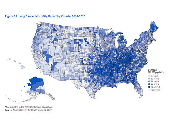
Lung cancer is by far the leading cause of cancer death among both men and women
in the United States. Our research program has played a role in many of the prevention, screening, and treatment advances that save lives from lung cancer today. And, we continue to fund research to help save even more lives in the future.
Current Status - In Brief
Survival After a Diagnosis of Lung Cancer Has Improved, Yet More People Die from Lung Cancer Than Any Other Type
Over the past 20 years, there have been exciting improvements in survival after a diagnosis of lung cancer. These are due to advances in the understanding of tumor biology, the development of targeted treatment, and the introduction of screening. However, we've known about the link between smoking and lung cancer for even longer, and smoking is still responsible for most deaths from lung cancer.
Risk & Prevention Studies
Most E-cigarette Users Ages 18 to 29 Never Smoked Before
Researchers say about 2.7 million younger adults with no history of cigarette smoking used e-cigarettes in 2021.
This graphic is from a study about the use of e-cigarettes by adults in the United States between 2019 and 2021 by American Cancer Society researchers and published in the American Journal of Preventive Medicine.
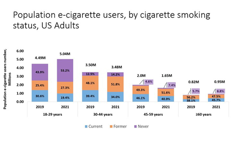
For 18 to 29-year-olds, the purple sections in 2019 and 2021 show that a large percentage of younger adults using e-cigarettes had never used traditional cigarettes. For all the other age ranges, the orange sections for both 2019 and 2021 show that the largest percentages of these populations using e-cigarettes had previously used traditional cigarettes.
Learn more about this graphic and the trends about the use of e-cigarette.
The ACS Tobacco Control Research Team Helps Put Evidence Into Action Via Policies
We study factors in the United States that predict what leads adults and adolescents to start and stop using any one of the many conventional or novel tobacco products. We track the use of cigarettes, cigars, and snuff along with electronic cigarettes (e-cigarettes), hookahs, and nicotine pouches. We also evaluate the value of tobacco control laws and rules—how effective they are in reducing the use of tobacco products in the US."

Screening & Early Detection Studies
New Lung Cancer Screening Guideline Increases Eligibility
The new lung cancer screening guideline, published in the ACS flagship journal, CA: A Cancer Journal for Clinicians, recommends that primary care or specialty care providers refer 50 to 80-year-olds for yearly screening with LDCT if they currently smoke or used to smoke, have a 20-pack-year or more smoking history, and are in reasonably good health, without any symptoms of lung cancer.
How does the new guideline differ from the previously published guideline?

Treatment Studies
Mapping Mitochondria's “Dance” May Be a Path to Starving NSCLC
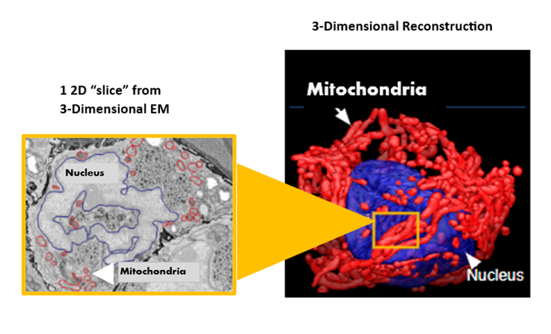
The image on the left is a flat, 2D image from an electronic microscope, with mitochondria circled by hand. The software's analysis generated a 3D image of mitochondrial networks in NSCLCs (on the right). The nucleus of a lung cancer adenocarcinoma cell is blue, and the mitochondrial networks are red.
The left image above shows manual labeling. A post-doctoral researcher traced each mitochondrion on a flat image from an electronic microscope. It took her more than 2 weeks to mark up hundreds of images.
In contrast, the scientists' deep-learning software analyzed 200 to 500 serial 2D images from the electron microscope, with 20,000 to 50,000 mitochondria within each tumor section in about 4 hours to create 3D images like the one shown on the right below.
Read more about how mitochondria move in different patterns to keep "feeding" a growing tumor.
Glossary for Nonscientists
Featured Term:
Deep-learning
Survivorship Studies
Noteworthy Statistics About Lung Cancer
Full Reports on Lung Cancer Statistics & Facts
We Fund Cancer Researchers Across the US
The American Cancer Society funds scientists who conduct research about cancer at medical schools, universities, research institutes, and hospitals throughout the United States. We use a rigorous and independent peer review process to select the most innovative research projects proposals to fund.
Stats on the left are as of Aug. 1, 2023.
